38 diagram of the human eye without labels
Human Ear Diagram - Bodytomy The Structure of Human Ear. Helix: It is the prominent outer rim of the external ear. Antihelix: It is the cartilage curve that is situated parallel to the helix. Crus of the Helix: It is the landmark of the outer ear, situated right above the pointy protrusion known as the tragus. Auditory Ossicles: The three small bones in the middle ear ... Human eye - Wikipedia The human eye is a sensory organ, ... Diagram of a human eye (horizontal section of the right eye) 1. Lens, 2. Zonule of Zinn or Ciliary zonule, 3. Posterior chamber and 4. Anterior chamber with 5. ... Right eye without labels (horizontal section) The structures of the eye labeled
File:Diagram of human eye without labels.svg - Wikimedia Commons File:Diagram of human eye without labels.svg. Size of this PNG preview of this SVG file: 410 × 430 pixels. Other resolutions: 229 × 240 pixels | 458 × 480 pixels | 732 × 768 pixels | 976 × 1,024 pixels | 1,953 × 2,048 pixels.

Diagram of the human eye without labels
Eye anatomy: A closer look at the parts of the eye In a number of ways, the human eye works much like a digital camera: Light is focused primarily by the cornea — the clear front surface of the eye, which acts like a camera lens. The iris of the eye functions like the diaphragm of a camera, controlling the amount of light reaching the back of the eye by automatically adjusting the size of the ... Eye Diagram - Differentiated Worksheets and EASEL Activities - Pinterest Eye Diagram - Differentiated Worksheets and EASEL Activities Description Use these simple eye diagrams to help students learn about the human eye. Three differentiated worksheets are included: 1. Write the words using a word bank 2. Cut and paste the words 3. Anatomy of the eye: Quizzes and diagrams - Kenhub Take a look at the diagram of the eyeball above. Here you can see all of the main structures in this area. Spend some time reviewing the name and location of each one, then try to label the eye yourself - without peeking! - using the eye diagram (blank) below. Unlabeled diagram of the eye
Diagram of the human eye without labels. PDF Eye Anatomy Handout - National Eye Institute of light entering the eye. Lens: The lens is a clear part of the eye behind the iris that helps to focus light, or an image, on the retina. Macula: The macula is the small, sensitive area of the retina that gives central vision. It is located in the center of the retina. Optic nerve: The optic nerve is the largest sensory nerve of the eye. Human Eye Ball Anatomy & Physiology Diagram - eMedicineHealth Orbit. The orbit is the bony eye socket of the skull. The orbit is formed by the cheekbone, the forehead, the temple, and the side of the nose. The eye is cushioned within the orbit by pads of fat. In addition to the eyeball itself, the orbit contains the muscles that move the eye, blood vessels, and nerves. Identifying Medical Diagnoses and Treatable Diseases by Image ... - Cell Feb 22, 2018 · By employing a transfer learning algorithm, our model demonstrated competitive performance of OCT image analysis without the need for a highly specialized deep-learning machine and without a database of millions of example images (STAR Methods). Moreover, the model’s performance in diagnosing retinal OCT images was comparable to that of human ... Eyes - Layers of Learning | Human eye diagram, Parts of the eye, Eye ... Elementary Science. Description Use these simple eye diagrams to help students learn about the human eye. Three differentiated worksheets are included: 1. Write the words using a word bank 2. Cut and paste the words 3. Write the words without a word bank Labels include: eyebrow, eyelid, eyelashes, pupil, iris, and sclera.
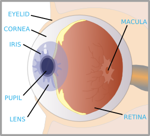
Label the Eye Diagram - Enchanted Learning Label the Eye Diagram. Human Anatomy. Read the definitions, then label the eye anatomy diagram below. Cornea - the clear, dome-shaped tissue covering the front of the eye. Iris - the colored part of the eye - it controls the amount of light that enters the eye by changing the size of the pupil. Lens - a crystalline structure located just behind ... Eye Anatomy: Parts of the Eye and How We See Behind the anterior chamber is the eye's iris (the colored part of the eye) and the dark hole in the middle called the pupil. Muscles in the iris dilate (widen) or constrict (narrow) the pupil to control the amount of light reaching the back of the eye. Directly behind the pupil sits the lens. The lens focuses light toward the back of the eye. Eye Diagram Unlabelled - schematron.org Best Human eye diagram unlabelled free vector download for commercial use in ai, eps, cdr, svg vector illustration graphic art design format. human eye. Ask A Biologistcoloring page | Web address:schematron.org coloring. Human Eye. Page 2. 5. 3. 2. 4. How to draw human eye in easy steps -10th -Physics - science - CBSE syllabus - NCERT class 10 Human penis - Wikipedia The human penis is an external male intromittent organ that additionally serves as the urinal duct.The main parts are the root (radix); the body (corpus); and the epithelium of the penis including the shaft skin and the foreskin (prepuce) covering the glans penis.The body of the penis is made up of three columns of tissue: two corpora cavernosa on the dorsal side and corpus …
File:Schematic diagram of the human eye en.svg - Wikimedia Diagram of the human eye in English. It shows the lower part of the right eye after a central and horizontal section. ... Full redraw: Group labels in accordance with the "Foundational Model Explorer." Added "Macula" and "Uvea" and removed "Zonular fibres." ... File:Diagram of human eye without labels.svg; File:Figure of diplopia perception ... Label the Eye Worksheet - Teacher-Made Learning Resources - Twinkl The first page is a labelling exercise with two diagrams of the human eye. One is a view from the outside, and the other is a more detailed cross-section. On the second page, you'll find a set of answers showing the properly labelled human eyes, designed to help you check the worksheets without having to come up with your own answer key. Anatomy of the Eye | Johns Hopkins Medicine The back part of the eye's interior. Pupil. The opening in the middle of the iris through which light passes to the back of the eye. Retina. The light-sensitive nerve layer that lines the inside of the back of the eye. The retina senses light and creates impulses that are sent through the optic nerve to the brain. Sclera. Structure and Functions of Human Eye with labelled Diagram The human eyes are the most complicated sense organs in the human body. From the muscles and tissues to nerves and blood vessels, every part of the human eye is responsible for a certain action. Furthermore, contrary to popular belief, the eye is not perfectly spherical; instead, it is two separate segments fused together.
Eye Model Labeled - Bing Images | Anatomy | Pinterest
Parts of Stereo Microscope (Dissecting microscope) – labeled diagram ... The human brain is able to generate a 3-D image through the optical path and the angular offset. CMO or “Parallel style” stereo microscopes do not have double lenses; instead, the microscope has only one objective lens with a large diameter through which the light paths run for both the left and the right eye.
How Vision Works: Our Sense of Sight | Ask A Biologist
Blank Eye Diagram - Healthiack Best viewed on 1280 x 768 px resolution in any modern browser. Blank eye diagram 1063. Blank eye diagram 1020. Blank eye diagram 1023. Blank eye diagram 1029. Blank eye diagram 1031. Blank eye diagram 1033. Blank eye diagram 1034. Blank eye diagram 1035.

picture front of the eye without labels clipart - Clipground
Human Body Diagram - Bodytomy ☛ The human eye has the ability to differentiate between 400+ shades of gray, and what's more, it can identify approximately 10 million colors. ☛ Your ears never sleep. Sound is received even while you are asleep; it's the brain that does not process them.

picture front of the eye without labels clipart - Clipground
Eye Anatomy Detail Picture Image on MedicineNet.com Picture of Eye Anatomy Detail. The eye is our organ of sight. The eye has a number of components which include but are not limited to the cornea, iris, pupil, lens, retina, macula, optic nerve, choroid and vitreous. Cornea: clear front window of the eye that transmits and focuses light into the eye. Iris: colored part of the eye that helps ...

How the eye works - Medical Information Illustrated
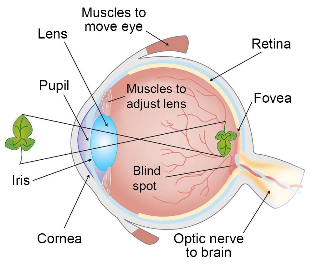
PDF Parts of the Eye - National Eye Institute | National Eye Institute Eye Diagram Handout Author: National Eye Health Education Program of the National Eye Institute, National Institutes of Health Subject: Handout illustrating parts of the eye Keywords: parts of the eye, eye diagram, vitreous gel, iris, cornea, pupil, lens, optic nerve, macula, retina Created Date: 12/16/2011 12:39:09 PM

13 best Eye Diagrams images on Pinterest | Eyes, Eye anatomy and Human anatomy
Human Figure Png - Human Body Diagram Without Labels PNG ... - SeekPNG.com Human Figure Png - Human Body Diagram Without Labels It is a very clean transparent background image and its resolution is 763x2315 , please mark the image source when quoting it. Seeking more PNG image human brain png,human png,human eyes png?
Eye With Labels Clip Art at Clker.com - vector clip art online, royalty free & public domain
Cornea of the Eye - Definition and Detailed Illustration Cornea Definition. The cornea is the clear front surface of the eye. It lies directly in front of the iris and pupil, and it allows light to enter the eye. Viewed from the front of the eye, the cornea appears slightly wider than it is tall. This is because the sclera (the "white" of the eye) slightly overlaps the top and bottom of the anterior ...

The Eye - Science Quiz
The Eyes (Human Anatomy): Diagram, Optic Nerve, Iris, Cornea ... - WebMD Your eye is a slightly asymmetrical globe, about an inch in diameter. The front part (what you see in the mirror) includes: Iris: the colored part. Cornea: a clear dome over the iris. Pupil: the ...

Eye Diagram Blank - Human Anatomy
Eye Diagram Teaching Resources | Teachers Pay Teachers Anatomy of the Eye Diagrams for Coloring/Labeling, with Reference and Summary by Homemade For Play 7 $1.95 PDF This printable contains 13 clear and simple cross sectional diagrams of the human eye.
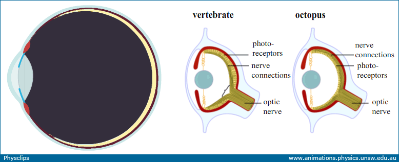
Eye:optics, anatomy and accommodation: Physclips - Light
Label Parts of the Human Eye - University of Dayton Label Parts of the Human Eye. Select One Anterior Chamber Ciliary Body Cornea Fibrous Tunic Iris Lateral Rectus Muscle Lens Medial Rectus Muscle Optic Disk Optic Nerve Pupil Retina Vascular Tunic Vitreous Nerve. Select One Anterior Chamber Ciliary Body Cornea Fibrous Tunic Iris Lateral Rectus Muscle Lens Medial Rectus Muscle Optic Disk Optic ...
parts of the eyes clipart - Clipground
Eye Anatomy: Parts of the Human Eye - Vision Center The lens of the eye (or crystalline lens) is the transparent lentil-shaped structure inside your eye. This is the natural lens. It is located behind the iris and to the front of the vitreous humor (vitreous body). The vitreous humor is a clear, colorless, gelatinous mass that fills the gap between the lens and the retina in the eye.

Human Eye Diagram To Label Ks2 - Food Ideas
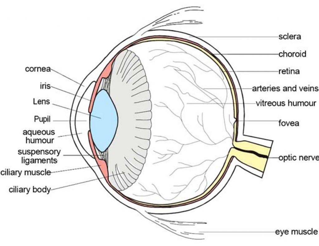
Eye Diagram With Labels and detailed description - BYJUS A brief description of the eye along with a well-labelled diagram is given below for reference. Well-Labelled Diagram of Eye The anterior chamber of the eye is the space between the cornea and the iris and is filled with a lubricating fluid, aqueous humour. The vascular layer of the eye, known as the choroid contains the connective tissue.

File:Schematic diagram of the human eye is.svg - Wikimedia Commons
Anatomy of the eye: Quizzes and diagrams - Kenhub Take a look at the diagram of the eyeball above. Here you can see all of the main structures in this area. Spend some time reviewing the name and location of each one, then try to label the eye yourself - without peeking! - using the eye diagram (blank) below. Unlabeled diagram of the eye

Labeled Diagram Of An Eye - Eye Anatomy A Closer Look At The Parts Of The Eye - Just the ...
Eye Diagram - Differentiated Worksheets and EASEL Activities - Pinterest Eye Diagram - Differentiated Worksheets and EASEL Activities Description Use these simple eye diagrams to help students learn about the human eye. Three differentiated worksheets are included: 1. Write the words using a word bank 2. Cut and paste the words 3.

Label Parts of the Human Ear
Eye anatomy: A closer look at the parts of the eye In a number of ways, the human eye works much like a digital camera: Light is focused primarily by the cornea — the clear front surface of the eye, which acts like a camera lens. The iris of the eye functions like the diaphragm of a camera, controlling the amount of light reaching the back of the eye by automatically adjusting the size of the ...
Schematic Diagram Eye Human Anatomy Labeled Stock Illustration 298561235 - Shutterstock

1000+ images about The Human Eye on Pinterest
Post a Comment for "38 diagram of the human eye without labels"