39 brain mri with labels
› doi › 10A unified 3D map of microscopic architecture and MRI of the ... Apr 27, 2022 · The inclusion of five microscopy labels, blockface images, and three quantitative MRI contrasts provides a wealth of anatomical information ().The full-brain coverage allows for detailed and comparative analyses of architectonic features for mapping the cortical laminar structure (20–23). Political Orientations Are Correlated with Brain Structure in Young ... Apr 26, 2011 · Apart from the anterior cingulate cortex, other brain structures may also show patterns of neural activity that reflect political attitudes. Conservatives respond to threatening situations with more aggression than do liberals [] and are more sensitive to threatening facial expressions [].This heightened sensitivity to emotional faces suggests that individuals with …
A unified 3D map of microscopic architecture and MRI of the human brain Apr 27, 2022 · The inclusion of five microscopy labels, blockface images, and three quantitative MRI contrasts provides a wealth of anatomical information ().The full-brain coverage allows for detailed and comparative analyses of architectonic features for mapping the cortical laminar structure (20–23).A second important application is the atlasing of small brain structures that …
Brain mri with labels
Harvard University Show labels Show list All modalities to: MR-T1 MR-T2 FDG T1/FDG T2/FDG github.com › naldeborgh7575 › brain_segmentationGitHub - naldeborgh7575/brain_segmentation Jun 16, 2017 · The segmentation labels are represented as follows: Figure 1: Ground truth segmentation overlay on a T2 weighted scan. MRI Background. Magnetic Resonance Imaging (MRI) is the most common diagnostic tool brain tumors due primarily to it's noninvasive nature and ability to image diverse tissue types and physiological processes. Brain Tumor Prediction Through MRI Images Using CNN In Keras Aug 19, 2020 · Both the folders contain different MRI images of the patients. Yes folder has patients that have brain tumors whereas No folder has MRI images of patients with no brain tumor. There are a total of 155 images of positive patients of brain tumor and 98 images of other patients having no brain tumor. All the images are of 240X240 pixels.
Brain mri with labels. volBrain: Automated MRI Brain volumetry system volBrain is an online MRI brain volumetry system. It is intended to help researchers all over the world to obtain automatically volumetric brain information from their MRI data without the need for any infrastructure in their local sites. ... Comparing fully automated state-of-the-art cerebellum parcellation from Magnetic Resonance Imaging ... › pmc › articlesMRI Segmentation of the Human Brain: Challenges, Methods, and ... Mar 01, 2015 · An example of the brain MRI segmentation with an original MR image (a) and segmented image with three labels: WM, GM, and CSF (b). Image segmentation can be performed on 2D images, sequences of 2D images, or 3D volumetric imagery. GitHub - naldeborgh7575/brain_segmentation Jun 16, 2017 · The segmentation labels are represented as follows: Figure 1: Ground truth segmentation overlay on a T2 weighted scan. MRI Background. Magnetic Resonance Imaging (MRI) is the most common diagnostic tool brain tumors due primarily to it's noninvasive nature and ability to image diverse tissue types and physiological processes. analyticsindiamag.com › brain-tumor-predictionBrain Tumor Prediction Through MRI Images Using CNN In Keras Aug 19, 2020 · Both the folders contain different MRI images of the patients. Yes folder has patients that have brain tumors whereas No folder has MRI images of patients with no brain tumor. There are a total of 155 images of positive patients of brain tumor and 98 images of other patients having no brain tumor. All the images are of 240X240 pixels.
iseg2019.web.unc.edu › dataData | MICCAI Grand Challenge on 6-month Infant Brain MRI ... Training Dataset. One zip file with training images and manual labels is available for downloading. They were randomly chosen from Multi-visit Advanced Pediatric (MAP) Brain Imaging Study, which is the pilot study of Baby Connectome Project (BCP), with the following imaging parameters: CPT Code for MRI Brain, Breast, Lumbar Spine and Shoulder Find below the latest Radiology CPT codes for for MRI of Brain, Breast, Lumbar Spine and Shoulder: CPT Codes for MRI Lumbar spine In human Lumbar spine is represented by the 5 vertebrae in between the ribcage and the pelvis forming the largest segment of the vertebral column. Depending on the condition that one is treated on these parts of the ... Diffusion MRI - Wikipedia Diffusion-weighted magnetic resonance imaging (DWI or DW-MRI) is the use of specific MRI sequences as well as software that generates images from the resulting data that uses the diffusion of water molecules to generate contrast in MR images. It allows the mapping of the diffusion process of molecules, mainly water, in biological tissues, in vivo and non-invasively. MRI Segmentation of the Human Brain: Challenges, Methods, … Mar 01, 2015 · An example of the brain MRI segmentation with an original MR image (a) and segmented image with three labels: WM, GM, and CSF (b). Image segmentation can be performed on 2D images, sequences of 2D images, or 3D volumetric imagery.
› 2013 › 07CPT Code for MRI Brain, Breast, Lumbar Spine and Shoulder Find below the latest Radiology CPT codes for for MRI of Brain, Breast, Lumbar Spine and Shoulder: CPT Codes for MRI Lumbar spine In human Lumbar spine is represented by the 5 vertebrae in between the ribcage and the pelvis forming the largest segment of the vertebral column. Depending on the condition that one is treated on these parts of the ... › AANLIB › casesHarvard University Show labels Show list All modalities to: MR-T1 MR-T2 FDG T1/FDG T2/FDG Data | MICCAI Grand Challenge on 6-month Infant Brain MRI … Training Dataset. One zip file with training images and manual labels is available for downloading. They were randomly chosen from Multi-visit Advanced Pediatric (MAP) Brain Imaging Study, which is the pilot study of Baby Connectome Project (BCP), with the following imaging parameters:T1-weighted MR images were acquired with 144 sagittal slices: TR/TE = … Brain Tumor Prediction Through MRI Images Using CNN In Keras Aug 19, 2020 · Both the folders contain different MRI images of the patients. Yes folder has patients that have brain tumors whereas No folder has MRI images of patients with no brain tumor. There are a total of 155 images of positive patients of brain tumor and 98 images of other patients having no brain tumor. All the images are of 240X240 pixels.
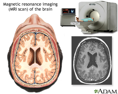
MRI of the brain: MedlinePlus Medical Encyclopedia Image
github.com › naldeborgh7575 › brain_segmentationGitHub - naldeborgh7575/brain_segmentation Jun 16, 2017 · The segmentation labels are represented as follows: Figure 1: Ground truth segmentation overlay on a T2 weighted scan. MRI Background. Magnetic Resonance Imaging (MRI) is the most common diagnostic tool brain tumors due primarily to it's noninvasive nature and ability to image diverse tissue types and physiological processes.

Radiodiagnosis - Imaging is Amazing-Interesting cases: January 2011
Harvard University Show labels Show list All modalities to: MR-T1 MR-T2 FDG T1/FDG T2/FDG

ON - RADIOLOGY: Lipoma arborescens of Ankle MRI (what`s that !!!)
MRI Procedure of Brain

RADIOLOGY: HIV Encephalopathy
Symptoms & Diagnosis

Neuroradiology Cases: Brain Abscess MRI

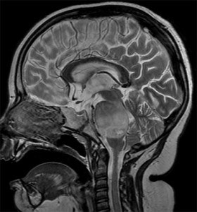
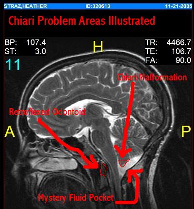
Annotated Sagittal T1 Midline MRI Scan of Reigh's Brain | Flickr
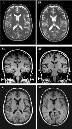
Brain MRI Comparison Images

Multiple sclerosis | Image | Radiopaedia.org

Bl4ck D4ys: March 2011
Images from Radiological Testing -- refried.org weblog

Brain MRI - NeurologyNeeds.com

Magnetic Resonance Imaging (MRI) Program - South Suburban College
Is it harmful brain MRI for human health

Membership Benefits And Executive Health For Everyone | Q Bio
Post a Comment for "39 brain mri with labels"