42 diagram of the brain without labels
Diagram Of Brain with their Labelings and Detailed Explanation The midbrain is the smallest region of the brain, found at the centre of the brain, between cerebral cortex and hindbrain. It comprises tectum, cerebral peduncle, tegmentum, cerebral aqueduct, substantia nigra, several nuclei and fasciculi. The midbrain is responsible for hearing, vision, sleep cycle, temperature regulation, alertness, etc. Picture of the Brain - WebMD • The cortex is the outermost layer of brain cells. Thinking and voluntary movements begin in the cortex. • The brain stem is between the spinal cord and the rest of the brain. Basic functions like...
Neuron Diagram Unlabeled neuron, (1). axon, cell body, dendrites, nucleus, terminal. Unlabeled diagram of a motor neuron (try labeling: axon, dendrite, cell body, myelin, nodes of Ranvier, motor end plate).Read the definitions, then label the neuron diagram below. axon - the long extension of a neuron that carries nerve impulses away from the body of the cell.

Diagram of the brain without labels
Brain Chart Maker - 100+ stunning chart types — Vizzlo Brain Chart overview and examples. Highlight and label different areas of the human brain. Create high-quality charts, infographics, and business visualizations for free in seconds. ... Adjust the position of each label by simply dragging the highlighted area into the desired spot. Brain chart maker: key features. Custom colors; This vizzard ... Labeled Diagrams of the Human Brain You'll Want to Copy Now The average dimension of the adult human brain is 5.5 inches in width and 6.5 inches in length. The height of the human brain is about 3.6 inches and it weighs about 4 to 5 lbs at birth and 3 lbs in adults. The total surface area of the cerebral cortex is about 2,500 cm2 and when stretched, it will cover the area of a night table. Neuron Diagram Unlabeled - Wiring Diagram Pictures A simple unlabeled tactile diagram of a neuron. Tactile Neuron Diagram ( Unlabeled) by trynne is licensed under the Creative Commons.Neuron Anatomy Activity The parts of the neuron have been labeled. Your challenge is to write the correct name for each part and explain what it does.
Diagram of the brain without labels. Diagram of the Brain and its Functions - Bodytomy Given below is a labeled diagram showing the brain stem and its related structures. Brain Stem and Structures Cerebellum The word 'cerebellum' literally means little brain. It is the second largest part of the brain, and is located at the back, below the occipital lobe, beneath the cerebrum and behind the brain stem. Blank ear diagrams and quizzes: The fastest way to learn - Kenhub It helps you to memorize the names and their locations, which in turn will aid you to remember their functions. Below, you can download both the blank ear diagram to make some notes, and then try labeling the ear using the unlabeled ear diagram. Good luck! DOWNLOAD PDF WORKSHEET (BLANK) DOWNLOAD PDF WORKSHEET (LABELED) Human Body Organs Diagram Stock Photos and Images - Alamy Body meridians - Schematic diagram with main acupuncture meridians and their directions of flow. Lungs, heart, liver, stomach, tooth human body organ icon set, flat internal organs silhouette vector symbol collection illustration. Medical diagram showing of the mechanisms of appetite regulation. endocrine system. Human Brain Diagrams and Detailed Information - Innerbody Brain. The brain is one of the most complex and magnificent organs in the human body. Our brain gives us awareness of ourselves and of our environment, processing a constant stream of sensory data. It controls our muscle movements, the secretions of our glands, and even our breathing and internal temperature.
75,682 Brain Anatomy Stock Photos and Images - 123RF Labeled diagram with location and functions. Frontal, parietal, occipital and temporal lobe scheme for human, mammal and reptilian brain. 3d rendered medically accurate illustration of the hippocampus The nervous system in the human brain with bright background Human Brain detailed anatomy. Vector Medical illustration. Parts of the Brain Activity for Kids, Brain Diagram, and Worksheets for ... Their are 2 brain function worksheets where your student will learn about the different parts of the brain your child will learn about are: FRONTAL LOBES - The frontal lobes control voluntary movement such as reasoning, planning, parts of speech and movement, emotions, and problem-solving It is fully developed by age 10. A&P2 Lab 2 HW Flashcards | Quizlet Drag the labels onto the diagram to identify the origins of the cranial nerves (VII - XII). look at pic The accumulation of blood during an epidural or subdural hemorrhage creates debilitating pressure on the brain and, without help, death is imminent. Cross-sectional anatomy of the brain - e-Anatomy - IMAIOS Cross sectional anatomy: MRI of the brain. An MRI was performed on a healthy subject, with several acquisitions with different weightings: spin-echo T1, T2 and FLAIR, T2 gradient-echo, diffusion, and T1 after gadolinium injection. We obtained 24 axial slices of the normal brain. Data and DICOM images archived on our PACS (Picture Archiving and ...
Human Nervous System - Diagram - How It Works | Live Science It consists of the brain, spinal cord and the retinas of the eyes. The Peripheral Nervous System consists of sensory neurons, ganglia (clusters of neurons) and nerves that connect the central... How to Draw a Brain: 14 Steps (with Pictures) - wikiHow Sketch a narrow curve above the top line of the brain. This will give your drawing a sense of dimension. Place your pencil on one end of the oval and draw a line that curves over the top line. It should be about 1⁄2 inch (1.3 cm) above the original outline at its widest point. How to make a Brain Model - Human Body Science for Kids Brain model with labels The outer part of the brain is called the Cerebrum, which is divided into two hemispheres, by a central fissure. Each Hemisphere is split into the 4 lobes, as shown above. Under the Cerebrum are the Cerebellum and the Brain Stem. The brain can be divided up into six main areas: The frontal lobe Label Brain Diagram Printout - EnchantedLearning.com Label the Brain Anatomy Diagram The Brain Read the definitions below, then label the brain anatomy diagram. Cerebellum - the part of the brain below the back of the cerebrum. It regulates balance, posture, movement, and muscle coordination. Corpus Callosum - a large bundle of nerve fibers that connect the left and right cerebral hemispheres.
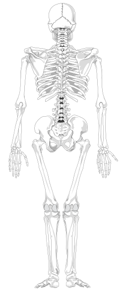
Human Skeleton Back No Text No Color Clip Art at Clker.com - vector clip art online, royalty ...
Label The Brain - Mr. Barth's Class You won't label the parts of the brain on this website, but you'll familiarize yourself with the location of the parts and their basic functions. Lobes of the Brain Click on the link to the left to label the lobes of the brain. See how quickly you can do it with 100% accuracy. Lobes and Neuron Diagram
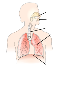
Unlabelled Respiratory System Clip Art at Clker.com - vector clip art online, royalty free ...
Skull Diagram Unlabeled - schematron.org Printable human skeleton diagram - labeled, unlabeled, and blank at skeleton. Human Skeleton Unlabeled The human body contains wood systems and organs which interact to keep both additional and interior problems of the body. The human body's maintenance system's importance should indeed be very high. Individuals can certainly get sick due.
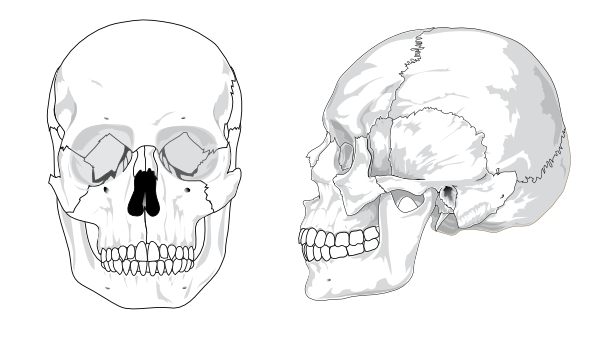
Human Skull No Text No Color Clip Art at Clker.com - vector clip art online, royalty free ...
Skull Anatomy - Cranial Bone and Suture Labeled Diagram, Names ... - EZmed The = Temporal Bones (2) Skull = Sphenoid Bone. This mnemonic not only helps you remember the cranial bone names, but also that there are 8 cranial bones (osseous parts) that form the skull. We are now going to discuss the anatomy and important features of each cranial bone in the order of the mnemonic. View fullsize.

Unlabeled Eye Diagram — UNTPIKAPPS
Anatomical diagrams of the brain - e-Anatomy - IMAIOS These original illustrations and diagrams of the brain were created from 3D medical imaging reconstructions and then redrawn and colored using Adobe Illustrator. These anatomical charts include the main diagrams necessary for medical students, nursing students, residents, practitioners, anatomists to study the anatomy of the brain, to ...

Microscope With Labels Clip Art at Clker.com - vector clip art online, royalty free & public domain
BYJUS BYJUS
Post a Comment for "42 diagram of the brain without labels"