39 lateral view of skull with labels
Identifying long-range synaptic inputs using genetically encoded labels ... Labeling was confirmed as reliable in this dataset as the mean pixel value of labeled mitochondria differed significantly from that of mitochondria contained in nearby putative unlabeled axons (N =... Anatomy Project - Sheridan College Neck. · Connecting the shaft and head of the femur. · Projects superior and medial from the shaft to the head. · In addition to projecting superior and medial from the shaft of the femur, the neck also projects somewhat anterior. · The amount of forward projection is extremely variable, but on an average is from 12° to 14°.
11 Possible Causes of Shooting Pain on Head - New Health Advisor Whenever you use medications, be sure that you are not taking more than the recommended dosage on the label. Other ways that you can release the tension headache is by applying a heating pad, taking a warm shower, getting some sleep, or getting a message to loosen the tense muscles. 3. Ice Pick Headaches

Lateral view of skull with labels
Elasmosaurus - Wikipedia Elasmosaurus (/ ɪ ˌ l æ z m ə ˈ s ɔːr ə s,-m oʊ-/;) is a genus of plesiosaur that lived in North America during the Campanian stage of the Late Cretaceous period, about 80.5 million years ago. The first specimen was discovered in 1867 near Fort Wallace, Kansas, US, and was sent to the American paleontologist Edward Drinker Cope, who named it E. platyurus in 1868. Anatomy And Physiology Archive | June 13, 2022 | Chegg.com Part A: Anatomy and Physiology in the Media - 7 points if both completed 1. Anatomy & Physiology in the News: (4 points) Find an article in a newspaper or magazine (in print or online) that discusses. 2. Anatomy & Physiology on TV: (3 points) Watch a TV show that deals with Human Anatomy & Physiology. Aorta: Anatomy, branches, supply - Kenhub The terminal branches of the abdominal aorta, the left and right common iliac arteries, arise from the bifurcation in front of the body of L4 vertebra about 1.25 cm to the left of the median plane. The common iliac arteries supply the lower limb, the gluteal region, and the pelvic viscera. Common iliac artery.
Lateral view of skull with labels. Posterior view of skeleton illustrations and clipart (183) Flat beige brown colour Vector illustration board Stock Illustration by Katya_Golovchyn 0 / 0 Upper limb Arm with Shoulder girdle Skeleton Human side lateral view. Anatomically correct realistic flat natural Clip Art by Katya_Golovchyn 0 / 0 Set of realistic skeletons isolated on gray background. Anterior, lateral and posterior view. Anatomy and relationships of the early diverging Crocodylomorphs ... Labels and details of these elements are shown in the following figures. Scale bar is equal to 5 cm. The holotype specimen consists of an exceptionally preserved skull and the anterior portion of the body, with a few disarticulated elements of the posterior portion of the skeleton that were recovered associated with the holotype (Figure 1 ). Horse Bones Blog by Swan Training The horse's body contains just over 200 bones, 205 to be exact. The alignment of these bones determines the horse's conformation, movement, mechanics, and efficiency. The bones of the horse skeleton are held together with ligaments, tendons, and muscles. When the skeleton structure is properly proportioned the joints work smoothly. Aberrant induction of p19Arf-mediated cellular senescence contributes ... Introduction. Cellular senescence is a form of permanent cell cycle arrest induced in response to a variety of stimuli. Senescence arrest is mediated by activation of cell cycle inhibitors including p21, p16 Ink4a, and p19 Arf [1-3].In addition, the arrested cells are highly secretory, producing a complex cocktail of cytokines, growth factors, extracellular matrix, and other proteins ...
Spinal Cord Cross Section Explained (with Videos ... - New Health Advisor In the center of the gray matter you will find the cerebrospinal fluid. The specific horns of the gray matter are responsible for different things. Each horn has different functions. The dorsal and ventral horns have the neurons that direct the skeletal muscles; the lateral horn has the cells that work the smooth muscle and supply cardiac. 3. Diagnostic captioning: a survey | SpringerLink Diagnostic captioning (DC) concerns the automatic generation of a diagnostic text from a set of medical images of a patient collected during an examination. DC can assist inexperienced physicians, reducing clinical errors. It can also help experienced physicians produce diagnostic reports faster. It is beneficial to invest resources to implement proton intracranial ... proton-based single or hypofractionated stereotactic radiosurgery (srs) for treating skull base meningiomas (>0.2 and <4.0 cm) has been reported decades ago using proton technology at the time, passive scattering with two beam arrangement. 2 - 4 excellent local control outcomes with these studies indicated the strong efficacy of proton-based srs … Nervous System: Anatomy, Structure, and Classification Sagittal Sagittal Computed Tomography (CT) view of the brain, brain stem Brain Stem The brain stem is a stalk-like structure that connects the cerebrum with the spinal cord and consists of the midbrain, pons, and medulla oblongata. It also plays a critical role in the control of cardiovascular and respiratory function, consciousness, and the sleep-wake cycle.
Clavicle: Anatomy and clinical notes - Kenhub The orientation of the clavicle can be distinguished by its ends: a broad, flat acromial end (referred to as the lateral third); and a round pyramidal-like sternal end (referred to as the medial two-thirds). Each end has unique bony landmarks, depending whether the superior or inferior surface of the bone is viewed. Superior surface How does Parkinson disease (PD) affect the basal ganglia ... - Medscape Answer. The basal ganglia motor circuit modulates the cortical output necessary for normal movement (see the following image). Schematic representation of the basal ganglia - thalamocortical motor ... Radiologic Technician Job Greenville Illinois USA,Healthcare Independently perform all radiographic procedures, i.e., chest, bone, skull, spines, and portable x-rays, PA and lateral, upper GI procedures, and barium enemas for the inmate population, etc. Notify Radiologist or referring Physician of any preliminary abnormalities seen while patient is undergoing an exam, so that additional views, or exams ... An Automated Deep Learning Model for the Cerebellum Segmentation from ... Cerebellum measures taken from routinely obtained ultrasound (US) images have been frequently employed to determine gestational age and identify developing central nervous system's anatomical abnormalities. Standardized cerebellar assessments from large-scale clinical datasets are required to investigate correlations between the growing cerebellum and postnatal neurodevelopmental results ...
Skull Quiz
Antenatal Care Module: 3. Anatomy and Physiology of the Female ... The vestibule is the area between the labia minora, and consists of the clitoris, urethral opening and the vaginal opening. The clitoris is a short erectile organ at the top of the vestibule, which has a very rich nerve supply and blood vessels. Its function is sexual excitation and it is very sensitive to touch.
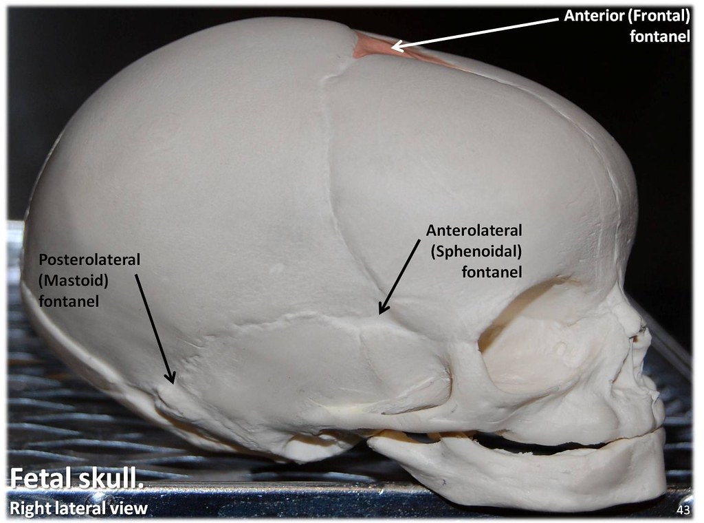
Fetal Skull, lateral view with labels - Axial Skeleton Visual Atlas, page 43 - a photo on Flickriver
Positions and Functions of the Four Brain Lobes - MD-Health.com The occipital lobe, the smallest of the four lobes of the brain, is located near the posterior region of the cerebral cortex, near the back of the skull. The occipital lobe is the primary visual processing center of the brain. Here are some other functions of the occipital lobe: Visual-spatial processing Movement and color recognition

Transcranial View of the Skull Base | Neuroanatomy | The Neurosurgical Atlas, by Aaron Cohen ...
Azerbaijan Medical Journal | AMJ Azerbaijan medical journal (ISSN: 0005-2523) - is a scopus indexed journal since 1961. The publisher of the journal is Izdatel'stvo Elm by WHO Office in Azerbaijan. Azerbaijan medical journal (AMJ) is also UGC approved. The journal publishes general medicine, health science, psychological, pharmaceutical journals and so on.

Skull diagram, lateral view with labels part 2 - Axial Ske… | Flickr
Dog - Wikipedia The skull, body, and limb proportions vary significantly between breeds, with dogs displaying more phenotypic diversity than can be found within the entire order of carnivores. ... A lateral view of a dog skeleton. All healthy dogs, ... showed that he knew the labels of over 200 different items. He inferred the names of novel things by ...

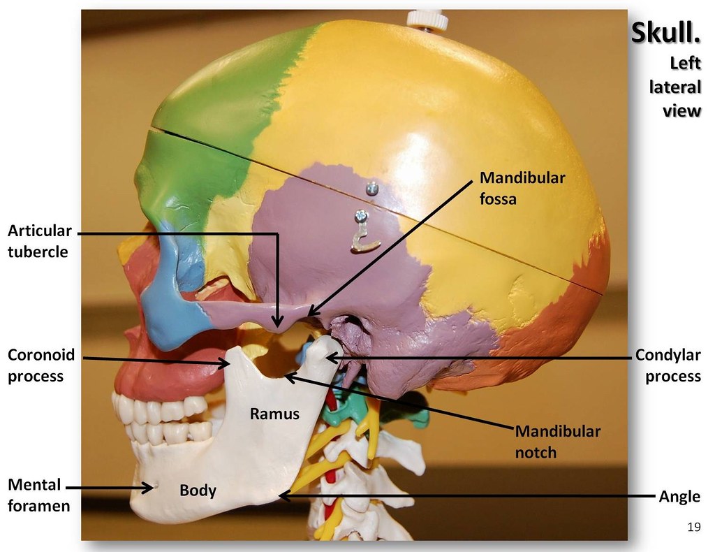
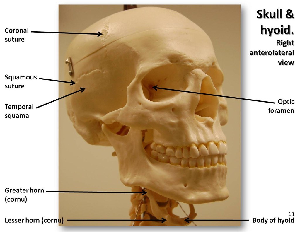
Lateral View of the Skull | Natalie Cormier
orbital floor fracture with entrapment - Zenobia Pulliam Lateral to the orbital canal lies the superior orbital fissure housing cranial nerves III IV V and VI. Orbital fracture with right eye entrapment. Example of a typical fracture involving the right orbital floor green arrow and medial maxillary sinus wall red arrow which is associated with resultant hemorrhage and an air-fluid level in the right ...

Lateral View of Skull | ClipArt ETC
Frontiers | Adenosine Downregulates the Activities of Glutamatergic ... Adult (8-12 weeks) vGlut2-Cre mice obtained from the Jackson lab (No. 016963). vGlut2-Cre mice were crossed with tdTomato reporter mice to label the glutamatergic neurons with the tdTomato in PVH. These adult transgenic mice were used to prepare the PVH slices. The coronal 400 μm slices containing the PVH were cut by a vibratome (7000SMZ, Campden).
Multi-colored Skull, lateral view with labels - Axial Skel… | Flickr
Aorta: Anatomy, branches, supply - Kenhub The terminal branches of the abdominal aorta, the left and right common iliac arteries, arise from the bifurcation in front of the body of L4 vertebra about 1.25 cm to the left of the median plane. The common iliac arteries supply the lower limb, the gluteal region, and the pelvic viscera. Common iliac artery.

Lesson 1.01: Context Clues
Anatomy And Physiology Archive | June 13, 2022 | Chegg.com Part A: Anatomy and Physiology in the Media - 7 points if both completed 1. Anatomy & Physiology in the News: (4 points) Find an article in a newspaper or magazine (in print or online) that discusses. 2. Anatomy & Physiology on TV: (3 points) Watch a TV show that deals with Human Anatomy & Physiology.

Dentistry lectures for MFDS/MJDF/NBDE/ORE: Diagrams Of Anatomy Of Skull With Radiographic Land Marks
Elasmosaurus - Wikipedia Elasmosaurus (/ ɪ ˌ l æ z m ə ˈ s ɔːr ə s,-m oʊ-/;) is a genus of plesiosaur that lived in North America during the Campanian stage of the Late Cretaceous period, about 80.5 million years ago. The first specimen was discovered in 1867 near Fort Wallace, Kansas, US, and was sent to the American paleontologist Edward Drinker Cope, who named it E. platyurus in 1868.

Anatomy Made Easy : Lateral View of Skull
Skull, anterolateral view with labels - Axial Skeleton Vis… | Flickr

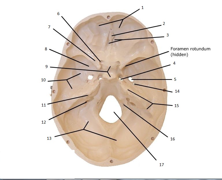
Inferior View of Skull Quiz (KNES 259)

Skull - Labeled Inferior View Photo by christinalimon81 | Photobucket

anatomyandphysiologytutor: “ A right lateral view of the skull, complete with labels ...
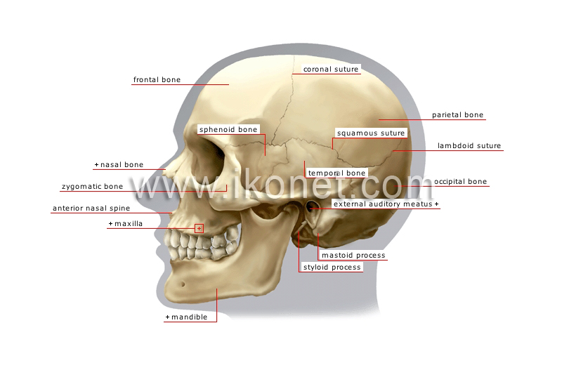
human being > anatomy > skeleton > lateral view of skull image - Visual Dictionary

Dentistry lectures for MFDS/MJDF/NBDE/ORE: Diagrams Of Anatomy Of Skull With Radiographic Land Marks
Post a Comment for "39 lateral view of skull with labels"